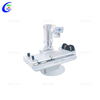
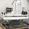
Dynamic(DR) DRF Digital X-Ray Photography System
Model number: MCI0729

Flat- panel detector
1.Detector type: Csl whole panel amorphous silicon flat detector
2.Detector panel size: 430mm*430mm
3.Detector effective imaging size: 17*17 inch
4.Detector acquisition pixel matrix: 3072*3072
5.AD conversion: 16 bits
6.Detector spatial resolution: photography 3.4LP / MM; fluoroscopy 2.4LP/ MM
7.Pixel gray scale: photography 16 Bit; fluoroscopy 14 Bit
8. Image acquisition and transmission time: 1.5s
High frequency and high voltage generator
1.Maximum output power: 65kW
2.Input power supply: 380VAC three- phase
3.Output voltage: 40~150kV
4.Tube current range: 10 ~ 800mA
5.perspective tube voltage: 40~125kV;
6.Continuous perspective tube current: 0.5-5mA
7.Current time accumulation range: 1mAs - 630mAs 8. Support AEC, APR automatic exposure
9. Failure self- diagnosis and display
X- ray tubule assembly
1.Focus: 0.6mm/1.2mm
2.Nominal electric power: 31kW / 74kW (150Hz)
3.Max. KV: 150kV
4.Anode target Angle: 12°
5.. Maximum heat dissipation rate of the anode: 475W
Upright column
1.Wave range of column: - 30°~30°, deviation is ± 2°
2.Horizontal movement range of column: 0~1000mm, deviation is ± 5mm
3.SID movement range: 1000~1800mm, deviation is ± 5mm
4.detector rotation angle range: - 45°~ +45°, deviation is ± 2°.
Electric Photography Fluoroscopy Table
1.Turning angle range of bed body: +90°~0°~ -20°; deviation ± 2
2.Horizontal movement range of bed surface; 250mm error ± 5°
3.Pressurizer pressure: 100N and 150N error ± 5N
4.Pressurize movement stroke is greater than 280mm, lowest point is less than 130mm, maximum pressure is less than 20kg
5.Tabletop height is less than 950 error ± 5mm
Grid and Beam limiter
1.Gate: aluminum grid
2.Size: 1818 inches (48*48cm)
3.Filter grid ratio is 10:1
4.Line / inch: 215C
5.Line grid focus: 1300mm
6.Light field, multi- blade DR special beam limiter
Image acquisition / processing and diagnostic workstation software function
1.Chinese interface, standard DICOM3.0 image
2.Image acquisition workstation function: adjust or preset window width /window position, local automatic window width / window position, preset window width / window position, positive and negative image flip, image flip, rotation, image amplification and roaming, image interpolation edge increase, strong, local amplification, restore the original image annotation, text / digital annotation, image marking, ruler segment measurement, square / circle measurement, arbitrary shape measurement, Angle measurement automatic electronic shear, image splicing, exposure index (Exposure Index) acquisition and display.
3.Special image acquisition and control software package, Special site protocol processing software package
4.With patient management, image acquisition, image processing (image correction, image flipping, USM sharpening, image filtering), image observation (providing image observation tools, Measurement tools), image splicing
5.Image printing: DICOM printing, paper printing, manual selection printing of displayed images, single key selection mark image printing, can select different printing equipment, film format and number of print sheets, print queue control, Stop / start pre-setting;
6. User personalization: display format and layout, default value setting, toolbar setting, site protocol enhancement filter
7.Image Display: Display configuration supports 19201080, high definition display.
8. Real- time preservation and playback of dynamic images.
WS
1.CPU: I79400
2.Host memory: 16GB
3.Hard disk: high capacity high speed hard disk 1T / 7200rpm
4.Workstation monitor: 24 " IPS LCD monitor
5.Communication network card and network interface: 1000M network card, 1000M network interface, Jumbo Frames: 9K
6.DICOM3.0 interface
7.Windows 10 64bit SP1 (Professional Edition or higher)
FAQ
1.What is the delivery time?
We have shipping agent,we can deliver the products to you by express,air freight,sea.Below is some delivery time for your reference: Express:UPS,DHL,TNT,ect (door to door) United States(3 days),Ghana(7 days),Uganda(7-10 days),Kenya(7-10 days),Nigeria(3-9 days) Hand carry Send to your hotel,your friends,your forwarder,your sea port or your warehouse in China. Air freight(from airport to airport) Los Angeles(2-7 days),Accra(7-10 days),Kampala(3-5 days),Lagos(3-5 days),Asuncion(3-10 days) Se
2.Technology R & D
We have a professional R&D team that continuously upgrades and innovates products.
3.What is your after-sales service?
We provide technical support through operating manual and video; Once you have questions, you can get our engineer's prompt response by email,phone call,or training in factory. If it's hardware problem, within the warranty period, we will send you spare parts for free, or you send it back then we repair for you freely.
Advantages
1.OEM/ODM, customized according to your requirements.
2.MeCan Focus on medical equipments over 15 years since 2006.
3.MeCan provide one-stop solutions for new hospitals,clinics,labs and universities,has helped 270 hospitals,540 clinics,190 vet clinics to set up in Malaysia,Africa,Europe,etc.we can save your time,energy and money.
4.MeCan offer professional service,our team is well-tained
About MeCan Medical
Guangzhou MeCan Medical Limited is a professional medical and laboratory equipment manufacturer and supplier. For more than ten years, we engage in supplying competitive price and quality products to many hospitals and clinics, research institutions and universities. We satisfy our customers by offering comprehensive support, purchase convenience and in time after sale service. Our main products include Ultrasound Machine, Hearing Aid, CPR Manikins, X-ray Machine and Accessories, Fiber and Video Endoscopy, ECG&EEG Machines,
Anesthesia Machines,
Ventilators,
Hospital furniture, Electric Surgical Unit, Operating Table, Surgical Lights,
Dental Chairs and Equipment, Ophthalmology and ENT Equipment, First Aid Equipment, Mortuary Refrigeration Units, Medical Veterinary Equipment.