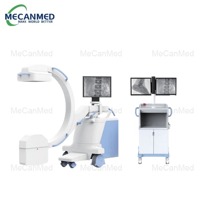
An X-ray machine is a diagnostic tool that uses electromagnetic radiation to create images of the inside of the body, allowing healthcare providers to examine bones, tissues, and organs for various medical conditions. Unlike other imaging methods, X-rays can penetrate the body and capture different densities, helping doctors visualize hidden areas. X-ray machines come in fixed or portable forms, with portable versions used for emergencies or bedside care. Understanding how an X-ray machine works is important for alleviating concerns about the procedure and its safety, ensuring patients and healthcare workers feel confident in its use and appreciate its role in effective healthcare.
What is X-Ray Technology?
What Are X-Rays?
X-rays are a form of electromagnetic radiation, similar to visible light but with much higher energy and shorter wavelengths. This allows X-rays to penetrate through different materials, such as the human body, and interact with tissues in distinct ways. The energy from X-rays passes through softer tissues and is absorbed by denser materials, such as bones, creating an image based on the amount of radiation that is transmitted through the body.
X-rays are typically generated by an X-ray tube, which accelerates electrons and directs them towards a target material (usually tungsten). The collision of electrons with the target material produces X-ray radiation, which is then used to capture images on film or digital sensors.
How X-Rays Differ from Other Types of Radiation
While X-rays are a form of ionizing radiation, they differ from other types of radiation like radio waves or microwaves. Ionizing radiation has enough energy to remove tightly bound electrons from atoms, which can potentially damage or alter living tissue. This makes the controlled use of X-rays important for safety. In comparison, radio waves and microwaves have much lower energy levels and are not capable of ionizing atoms, making them harmless in the context of medical imaging.
Components of an X-Ray Machine
What Are the Main Parts of an X-Ray Machine?
X-ray Tube: The X-ray tube is where X-rays are generated. It consists of a cathode (negative electrode) that emits electrons and an anode (positive electrode) that targets those electrons to produce X-rays. The tube operates in a vacuum to allow the electrons to travel unimpeded.
Control Panel: The control panel allows the operator to adjust settings like exposure time, intensity, and angle of the X-ray. This is essential for capturing clear and accurate images while minimizing radiation exposure.
Detector (Film or Digital Plate): After X-rays pass through the body, they hit the detector, which records the remaining radiation. Traditional X-rays used photographic film to capture images, but modern machines use digital detectors that provide clearer, more detailed images and are easier to store and share.
Collimator: A collimator is a device that shapes the X-ray beam to target the area of interest. This reduces unnecessary exposure to radiation in other parts of the body, improving safety.
Protective Lead Shields: Lead shields are used to protect sensitive areas of the body from radiation, such as the thyroid, reproductive organs, and eyes. These shields ensure that only the necessary areas are exposed to the X-rays.
How Do X-Ray Machines Produce Images?
The X-ray machine works by directing a beam of X-rays towards the patient’s body. As the X-rays pass through, some are absorbed by denser materials (like bones), and others pass through softer tissues. The radiation that passes through the body reaches the detector, where it is recorded. The varying levels of absorption create a shadow image of the body’s internal structure. Digital systems can process this data to generate highly detailed, often real-time images that are used for diagnosis.
The Process of Taking an X-Ray Image
How Does an X-Ray Machine Work in Practice?
To perform an X-ray, the patient is typically positioned between the X-ray tube and the detector. Depending on the area being imaged, patients may be asked to lie down, sit, or stand. The healthcare provider will adjust the X-ray machine’s angle and position to ensure the target area is properly aligned. The patient will then be asked to stay still for a few seconds while the image is captured. This brief exposure allows the X-ray beam to pass through the body and reach the detector.
What Happens After the X-Ray is Taken?
Once the X-ray is taken, the detector captures the image and sends it to a computer or film for processing. In traditional systems, the film is developed in a darkroom, but in digital systems, the images are displayed on a screen for immediate viewing. The processed images are reviewed by a radiologist or healthcare provider, who looks for signs of abnormalities or conditions like fractures, infections, or tumors.
Types of X-Ray Machines and Their Applications
What Are the Different Types of X-Ray Machines?
Fixed X-ray Machines: These are standard machines found in hospitals or clinics and are typically used for general radiography. They are permanently installed and offer high-resolution images.
Portable X-ray Machines: Smaller and mobile, portable X-ray machines are useful in emergency situations or for patients who cannot easily be transported to a fixed X-ray machine, such as those in intensive care units.
CT (Computed Tomography) Scanners: These machines use X-rays in combination with computer processing to create detailed cross-sectional images of the body, offering a 3D view. They are typically used for more complex imaging needs.
Fluoroscopy Machines: These provide real-time X-ray imaging and are commonly used in procedures such as catheter insertion, joint manipulation, and digestive tract imaging.
What Are the Common Medical Applications of X-Ray Machines?
Bone Fractures: X-rays are most commonly used to identify fractures in bones, whether from trauma or other causes.
Chest X-rays: These are frequently used to detect lung conditions such as pneumonia, tuberculosis, lung cancer, or heart enlargement.
Dental X-rays: Dentists use X-rays to examine the condition of teeth and gums, detect cavities, and plan treatments like root canals or implants.
Mammography: A specialized form of X-ray used for breast cancer screening. It can detect lumps or other abnormalities that may not be felt during a physical exam.

How Does an X-Ray Machine Work in Terms of Radiation Safety?
Is Radiation from X-Ray Machines Safe?
X-ray machines do expose the body to ionizing radiation, but the doses used in medical imaging are generally low. Radiation exposure is carefully controlled to minimize risks, and the benefits of diagnosing and treating medical conditions far outweigh the potential risks. X-ray technicians and radiologists take precautions to ensure that only the necessary area of the body is exposed to radiation, and they use the lowest effective dose to obtain clear images.
How Do Professionals Ensure Patient Safety During X-Ray Procedures?
Radiation safety during X-ray procedures is carefully managed through protocols like:
Positioning: Ensuring the patient is properly positioned to capture only the required area.
Lead Shields: Applying lead aprons or collars to shield vulnerable areas from radiation.
Minimizing Exposure: Using the minimum necessary exposure time to capture the image.
Monitoring: Regular checks of equipment to ensure proper function and safety.
Advances in X-Ray Technology
How Has X-Ray Technology Evolved Over the Years?
X-ray technology has evolved significantly since its invention in the late 19th century. From traditional film-based X-rays, we now have digital radiography, which offers higher image quality, quicker results, and easier sharing of images. Additionally, advancements like computed tomography (CT) and fluoroscopy have provided more detailed and dynamic imaging options. Modern systems also feature lower radiation doses, improving patient safety.
What Are the Future Trends in X-Ray Technology?
Future developments in X-ray technology include:
AI-Powered Imaging: AI and machine learning algorithms can assist in detecting abnormalities in X-ray images, making diagnoses quicker and more accurate.
Portable X-ray Systems: Smaller, lighter, and more flexible portable X-ray machines allow for more widespread use, particularly in emergency and remote settings.
Dose Reduction: Ongoing efforts to reduce radiation exposure while maintaining image quality, particularly for pediatric patients or those requiring frequent imaging.
Conclusion
X-ray machines are essential diagnostic tools that use electromagnetic radiation to create detailed images of the body’s internal structures, helping healthcare providers diagnose a wide range of medical conditions. Understanding how these machines work can ease patient concerns and reassure them about the safety of the procedure. With continuous advancements in technology, X-rays remain one of the most effective methods for diagnosing conditions, from fractures to life-threatening diseases like cancer. As technology progresses, X-ray systems continue to improve in precision and safety, offering even lower radiation exposure and enhancing overall patient care.
FAQ
Q: What is the Difference Between X-Ray and CT Scans?
A: X-rays provide 2D images, while CT scans create detailed 3D images using multiple X-ray slices.
Q: Are X-Rays Harmful to the Body?
A: X-rays use low radiation levels, and when used appropriately, they are safe with minimal risk.
Q: How Long Does an X-Ray Procedure Take?
A: Most X-ray procedures take just a few minutes, with the entire process often lasting under 15 minutes.
Q: Can I Have an X-Ray While Pregnant?
A: X-rays should be avoided during pregnancy unless medically necessary, as they may affect the fetus.
Q: How Often Can I Safely Get an X-Ray?
A: The frequency depends on medical need. Doctors minimize exposure and use the lowest effective dose.


