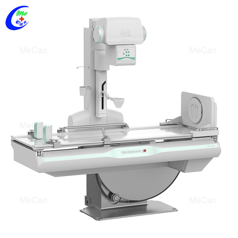
Professional Medical Dynamic FPD DRF Digital Radiography X Ray Machine factory
Model:MCX-6000

Features:
Equipped with the advanced pulse fluoroscopy technology, providing sharp image with low dose.
Intelligent Image Processing function of software offering high-quality image.
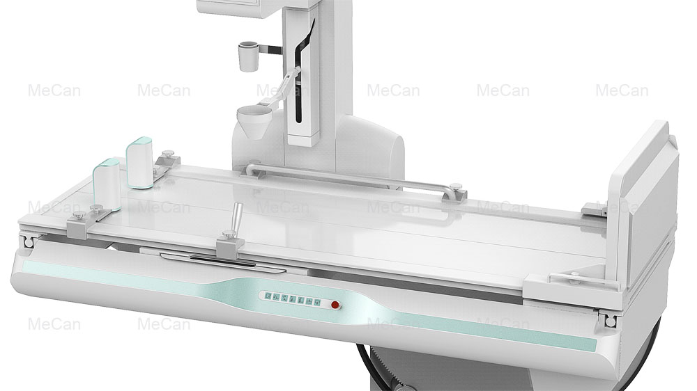
Flexible diagnostic table makes movement to meet all kinds of exposure needs.
Specification:
Item | Content | Technical parameter |
Power Supply | Voltage | 380V±38V |
frequency | 50Hz±1Hz |
capacity | ≥105kVA |
internal resistance | ≤0.17Ω |
High voltage generator (FSQ65RF) | Maximum output power | 65.5kW |
Main inverter frequency | 500kHz, tolerance ± 20% |
Radiography tube voltage | 40kV~150kV |
Radiography tube current | 10mA~800mA regulation in steps |
Radiography time | 1ms~ 10000ms regulation in steps |
Radiography mAs | 0.1~1000 mAs |
Fluoroscopy voltage | 40kV~ 125kV Continuously adjustable |
Fluoroscopy current | 0.5mA~10mA (Continuously fluoroscopy) 5mA~ 20mA (pulse fluoroscopy) |
Collimator (XSQ20) | Equivalent total filtration | ≥1mmAL |
beam limiter view light | LED lamp beads, DC3.6V/5W |
Visible light illumination and brightness | Center average irradiation brightness: > 100Lux |
Light and field exposure time | 5-45s, 5s per step |
X ray tube (E7252X) | Nominal anode input power | Big focus 75kW(180 Hz 0.1s) Small focus 27kW(180 Hz 0.1s) |
Anode heat capacity | 210KJ(300kHU) |
Component heat capacity | 475kJ(667kHU/min) |
Rotating anode speed | 9700rpm(180 Hz) |
Tube focus | Big focus 1.2mm /small focus 0.6mm |
Target angle | 12° |
Diagnostic (ZDC20F) | Table rotating range | +90 ° ~ 0 ° ~ -25 ° |
Longitudinal movement of point device | 1000mm, Tolerance ± 20 mm |
Lateral movement range of table surface | Not less than 220mm |
SID | 1000mm~1800mm;Tolerance ± 20 mm |
Rotating foot pedal | 360°Infinite rotation(optional) |
Filter grid | 498.5×499mm 230L/INCH 10:1 100cm 498.5×499mm 103L/INCH 10:1 180cm manual switching |
Dynamic flat panel detector (DRF-1717A) | Effective area | 434mm(H)×434mm(V) |
Prime matrix | 3072(H)×3072(V) |
Prime particle spacing | 139μm |
Pulse fluoroscopy | 12fps / 1408 x 1408 16fps / 1024 x 1024 22fps / 768x768 |
Continuous fluoroscopy | 13fps / 1408 x 1408 20fps / 1024 x 1024 30fps / 768x768 |
Serial radiography | 3fps / 3072 X 3072. |
6fps / 1536 X 1536. |
Point point | 3072 X 3072. |
1536 X 1536. |
Spatial resolution | ≥3.7lp/mm |
A / D Transformation | 16bit |
Energy range | 40 ~ 150 kVp |
Image output and control | Gigabit lan |
Image acquisition workstation:
1.Registration: routine registration, emergency registration, adding agreement, adding item, clearing information, starting inspection;
2.Work list: list information, patient search to be examined, refresh to be examined list, delete examination, display column settings. Start inspection and emergency registration;
3.Check list: list information, checked patient display and search, delete image, image storage, disc burning, add item, 4.display column setting, modify check information;
5.Patient's body type: thin adult, adult, fat adult;
6.Photography parameter setting: exposure mode, frame rate setting, kVp, Ma, MS, MAS, AEC, focus selection;
7.Perspective parameter setting: exposure mode, frame rate setting, kVp, Ma, ABS, time reset;
8.Browsing tools: zoom, horizontal flip, vertical flip, left turn 90 degrees, right turn 90 degrees, zoom in, zoom out, original size, moving image, reverse color, adaptive size, ROI magnifying glass, magnifying glass, default window width and position, window width and position of interest area, visual window width and position, point gray value, advanced processing, elliptical gray measurement;
9.System tools: text mark, front position mark, left mark, right mark, circle clipping, delete image, delete tool;
10.Error reset, exposure indication, full screen, save current image, print
11.Measurement tools: arrow, cardiothoracic ratio (CTR), distance measurement, angle measurement, spine measurement;
12.System tools: text mark, front position mark, left mark, right mark, circle clipping, delete image, delete tool;
13.Report, save current image, print
14.Report editing: patient information display and editing, photo image selection, report content template selection, report description, report conclusion, report description + conclusion, edit knowledge base, report doctor, audit doctor, report time, print template, setting and saving report;
15.Report printing: fast printing, printing report
Image archiving, burning, printing: delete image, image storage, browse image, report, lock / unlock, storage queue, print queue;
16.Disc recording: volume label, save settings, file compression, file structure;
17.Printing: DICOM printer, local printer
18.System settings: system, annotation information, tools, others;
19.Hardware configuration: syncbox, high voltage, detector, collimator, DAP;
20.Network configuration: local, worklist, netstore, local store, printing;
21.Inspection management: basic information, positioning information, hardware parameters, image processing, inspection protocol;
22.Quality management: search, system management;
23.User management: add, update, delete, permission.
more pictures of our x-ray machine:
FAQ
1.Quality Control (QC)
we have a professional quality control team to ensure that the final pass rate is 100%.
2.Technology R & D
We have a professional R&D team that continuously upgrades and innovates products.
3.What is the delivery time?
We have shipping agent,we can deliver the products to you by express,air freight,sea.Below is some delivery time for your reference: Express:UPS,DHL,TNT,ect (door to door) United States(3 days),Ghana(7 days),Uganda(7-10 days),Kenya(7-10 days),Nigeria(3-9 days) Hand carry Send to your hotel,your friends,your forwarder,your sea port or your warehouse in China. Air freight(from airport to airport) Los Angeles(2-7 days),Accra(7-10 days),Kampala(3-5 days),Lagos(3-5 days),Asuncion(3-10 days) Se
Advantages
1.MeCan provide one-stop solutions for new hospitals,clinics,labs and universities,has helped 270 hospitals,540 clinics,190 vet clinics to set up in Malaysia,Africa,Europe,etc.we can save your time,energy and money.
2.MeCan offer professional service,our team is well-tained
3.Every equipments from MeCan gets passed strict quality inspection,and final passed yield is 100%.
4.OEM/ODM, customized according to your requirements.
About MeCan Medical
Guangzhou MeCan Medical Limited is a professional medical and laboratory equipment manufacturer and supplier. For more than ten years, we engage in supplying competitive price and quality products to many hospitals and clinics, research institutions and universities. We satisfy our customers by offering comprehensive support, purchase convenience and in time after sale service. Our main products include Ultrasound Machine, Hearing Aid, CPR Manikins, X-ray Machine and Accessories, Fiber and Video Endoscopy, ECG&EEG Machines,
Anesthesia Machines,
Ventilators,
Hospital furniture, Electric Surgical Unit, Operating Table, Surgical Lights,
Dental Chairs and Equipment, Ophthalmology and ENT Equipment, First Aid Equipment, Mortuary Refrigeration Units, Medical Veterinary Equipment.