Introduction
Emergency and trauma care really is all about speed. Picture the chaos of an ER — paramedics burst through the doors, pushing a stretcher, that monitor screaming in the background. Trauma comes in fast, whether it’s a car crash or someone hurt on the job, and the team needs to figure out what’s wrong, right now. If they hesitate, even for a moment, things can get bad. Sometimes, those first few minutes decide everything — life or death, a full recovery or a permanent injury.
Take a patient who has been hurt in a serious car accident as an example. He/she might have hidden internal problems which are not visible on the surface, like brain bleeding, damage to organs (e.g., liver, spleen, etc,) or broken bones. Doctors need to quickly figure out how bad these injuries are so they can start the correct treatment.Without the proper equipment, they only can start the treatment relying on their own experience, which is not enough to save the patient’s life.
With its ability to create images quickly and accurately, the CT scan machine has become a vital tool in emergency and trauma care.


Fast Imaging: The Key to the Golden Hour
A core principle of trauma care is that "time is life." This is especially critical for patients with severe conditions like brain or multiple organ injuries. The ability to diagnose these injuries within the "golden hour" directly determines the effectiveness of treatment and the patient's ultimate outcome.
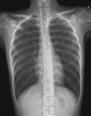
Although digital X-ray machines are simple to operate and can generate an image within 10 seconds, they can only provide a single flat image, which limits their diagnostic ability in complex trauma cases.


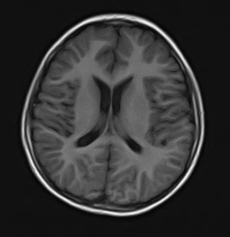
Although MRI machines boast superior clarity for soft tissue imaging and involves no ionizing radiation, their lengthy scan time — typically ranging from 15 minutes to over an hour — make it poorly suited for the time-sensitive demands of emergency and trauma care.


In the emergency and trauma care, CT scan machines address these limitations. Scanning one area only takes 5 to 15 seconds. Instead of a single flat image, CT scan machine provides detailed cross-sectional images, which offers a clearer view of internal structure. The short imaging time and clear image allows doctors to obtain a comprehensive diagnostic dataset immediately after a patient arrives, reducing the dangerous delays caused by repeated examinations.
When performing CT scan examination for a patient injured in a car accident, doctors can obtain images of full body in five to ten minutes. These images can help them immediately identify life-threatening injuries such as cerebral hemorrhage, pulmonary contusions, and lacerations of the internal organ, which is vital for doctors to implement target treatment (e.g., blood pressure control, respiratory function maintenance, emergency hemostasis).
Precision Targeting: Leaving No Hidden Injury Undetected
Traumatic injuries can be deeply deceptive: life-threatening damage can hide beneath a seemingly normal surface. Thus, these injuries demand exceptional diagnostic precision — even a minor mistake can mess up the treatment plan. This is where the CT scanner proves indispensable. Using millimeter-level imaging, it performs a virtual “dissection” of the body, building a detailed picture layer by layer, from the skin inward. Beyond simply detecting injuries, it locates them with accuracy, gauges their severity, and characterizes their nature — leaving no hidden threat overlooked.
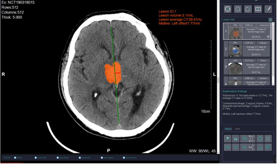
Consider the treatment of head trauma, for instance. While both epidural and intracerebral hematomas are types of intracranial hematomas, they actually differ in location, causes, symptoms, and treatment principle. Thus, making an accurate distinction between them are absolutely critical for treatment. The CT scan machine can provide precise localization and clear boundary images of each type of hematoma (the error margin of less than one millimeter). This precise imaging allows physicians to quickly assess the severity of the injury. It directly guides the critical decision between conservative management and emergency surgery, preventing any dangerous delays that stem from diagnostic uncertainty.

Differential Diagnosis: Finding the Answer in the Similar
In emergency and trauma care, many critical conditions often appear similar symptoms, which makes it difficult for doctors to differentiate the patient’s condition based on clinical presentation alone. A wrong diagnosis may make the subsequent treatment go in the wrong direction, which probably leads to severe consequences. With its ability to produce high-resolution images, the CT scan machine is vital to prevent the wrong diagnosis caused by similar symptoms. It can quickly help to get to the real cause behind similar symptoms, making sure doctors have what they need to treat patients the right way.
Take a pounding headache, for example. Sure, it’s often just a migraine, but sometimes it’s a warning sign for something far more serious — a brain bleed, a stroke, even a tumor. Strokes are a real headache (no pun intended) because they love to masquerade as each other. Ischemic or hemorrhagic, both can start with the same stuff: headache, a weak arm or leg, or suddenly losing coordination. It’s not always easy to spot the difference without the right evidence.

This is precisely where the CT scan machine steps in. In just a few seconds, it provides sharp, detail images of the brain. A hemorrhagic stroke will typically show up as a bright white area, while an ischemic stroke appears as a darker patch of compromised tissue. This obvious distinction allows physicians to spot the cause right away and initiate the correct, life-saving treatment — no waiting around.
Conclusion
In emergency and trauma care, the CT scan machine has become an indispensable heart of the action. Fast imaging buys precious time during the “golden hour.” Sharp, detailed images help doctors navigate even the trickiest injuries. Bringing CT technology into a hospital doesn’t just add a new machine. It raises the whole level of care. It gives doctors and nurses more to work with, and that means better chances for patients.
Enhancing Your Trauma Care with Advanced CT Imaging
This article has outlined the critical role of a CT scan machine in emergency and trauma care. Choosing the right one really matters. It can make all the difference for your patients. If you’re weighing your options, our imaging specialists are here to help. They’ll talk through your needs, break down the specs, and help you find the setup that fits your team and your workflow. And of course, everything stays confidential.


